

Muscles Labeled Front And Back Human Anatomy Body
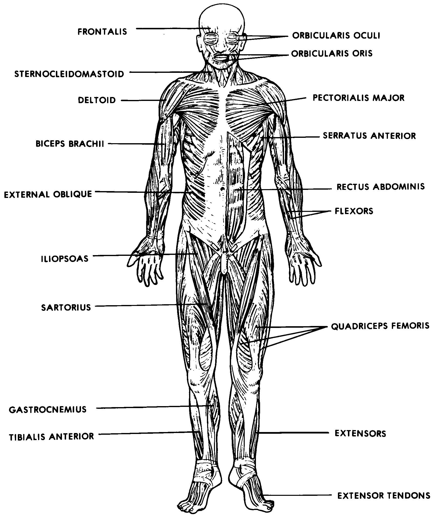
Human Anatomy - Front View of Muscles Click on the labels below to find out more about your muscles. More human anatomy diagrams: back view of muscles, skeleton, organs, nervous system Flex.
The Musculoskeletal System (Structure and Function) (Nursing) Part 4
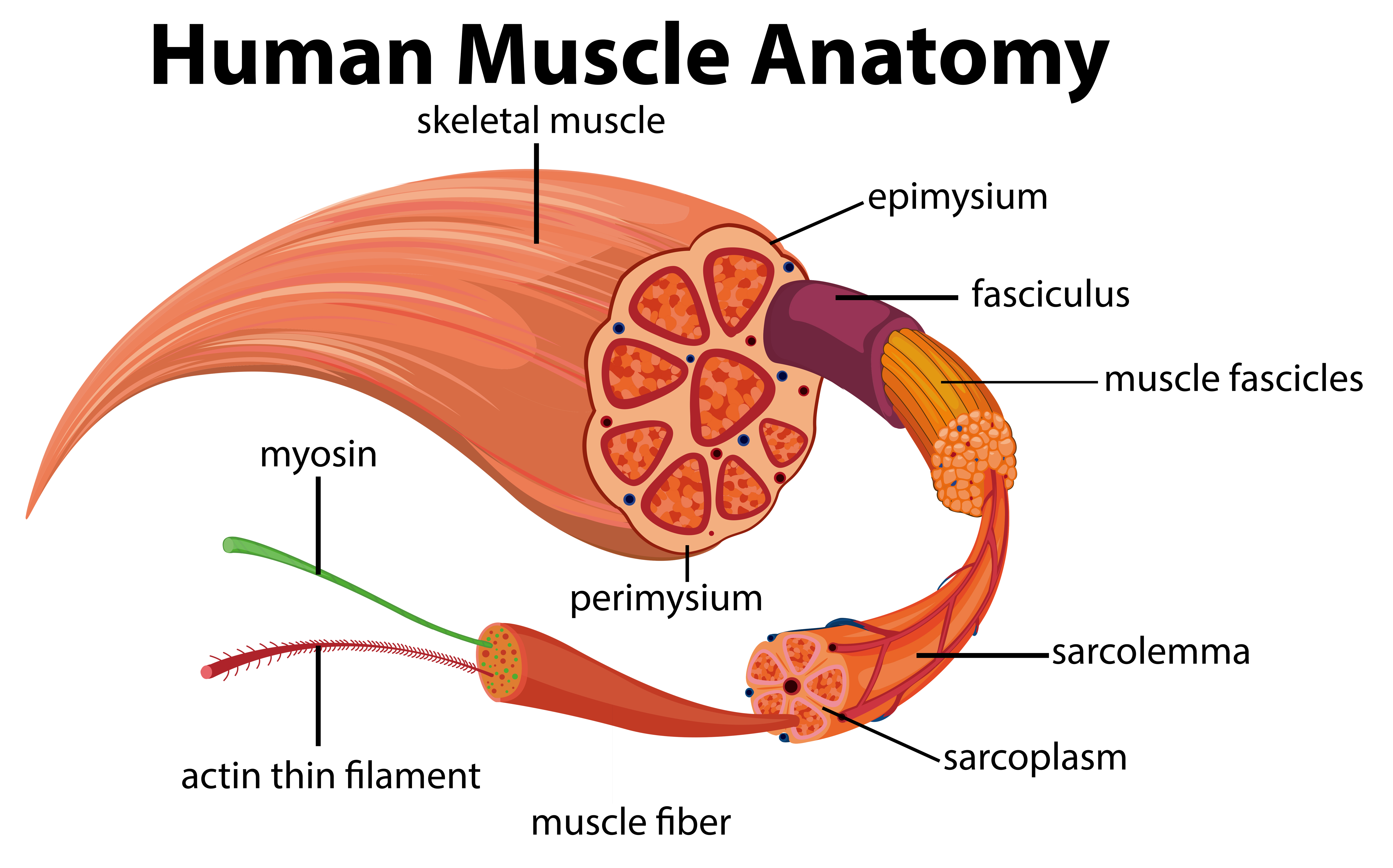
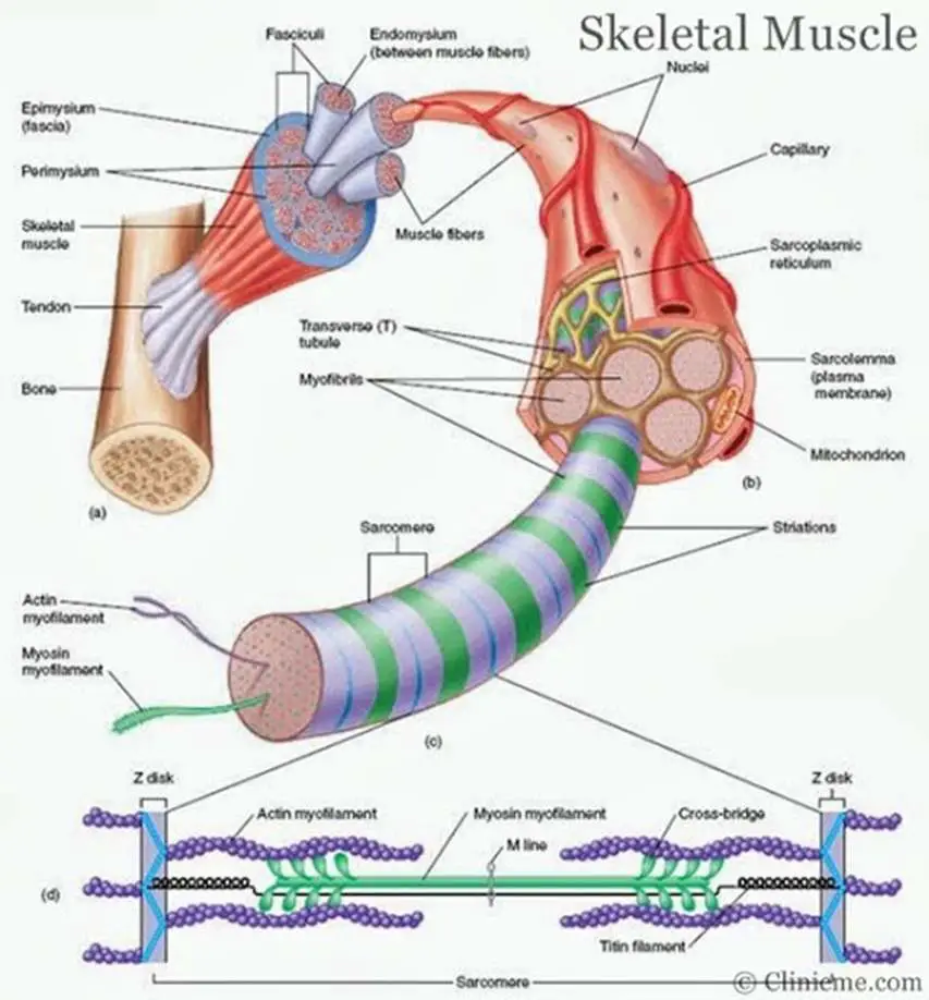
Muscles attach to bones directly or through tendons or aponeuroses. Skeletal muscles maintain posture, stabilize bones and joints, control internal movement, and generate heat. Skeletal muscle fibers are long, multinucleated cells. The membrane of the cell is the sarcolemma; the cytoplasm of the cell is the sarcoplasm.

Simple Human Muscles Diagram / Learn All Muscles With Quizzes And Labeled Diagrams Kenhub
Allow the barbell to lean your torso forward slightly. Squat by unlocking your hips and knees simultaneously, sinking downward as low as you're able. Maintain a braced core and a flat back as.
Human Muscle Anatomy Diagram 433295 Vector Art at Vecteezy
Solution Muscles: Muscular tissue is a specialized tissue in animals which applies forces to different parts of the body by contraction. There are three types of muscles such as skeletal muscle, smooth muscle, and cardiac muscle. Skeletal muscle: The striated muscle is another name for skeletal muscle.

10.2 Skeletal Muscle Anatomy & Physiology
Muscle structure Skeletal (striated or voluntary) muscle consists of densely packed groups of hugely elongated cells known as myofibers. These are grouped into bundles (fascicles). A typical myofiber is 2-3 centimeters ( 3/4-1 1/5 in) long and 0.05millimeters (1/500 inch) in diameter and is composed of narrower structures - myofibrils.

Labeled Muscle Diagram Chart Free Download
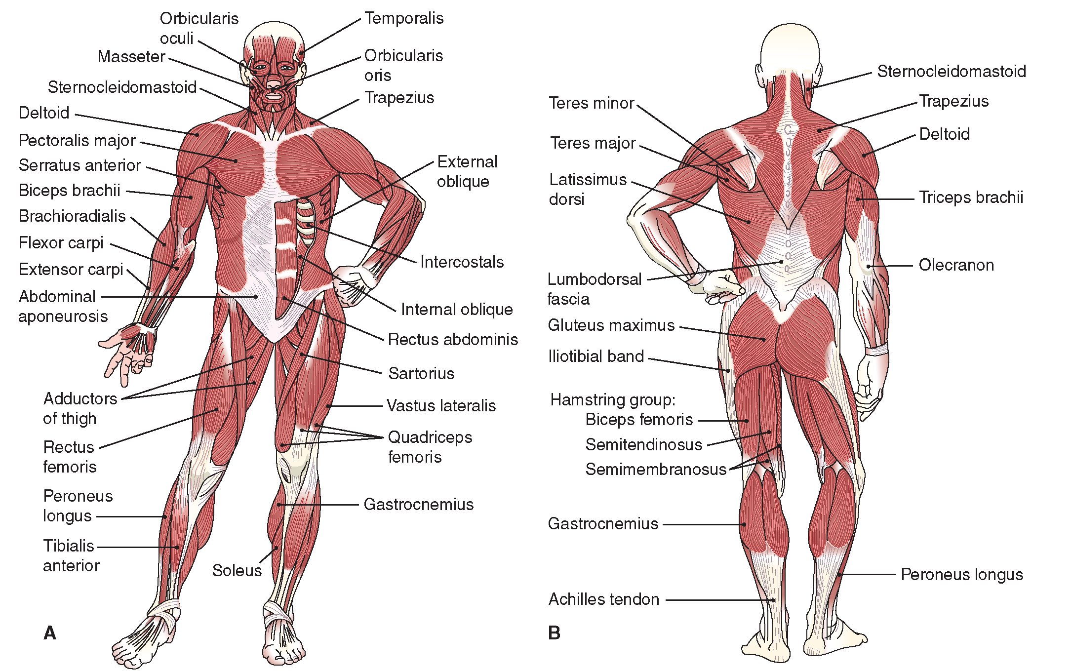
The muscular system is responsible for the movement of the human body. Attached to the bones of the skeletal system are about 700 named muscles that make up roughly half of a person's body weight. Each of these muscles is a discrete organ constructed of skeletal muscle tissue, blood vessels, tendons, and nerves.

Clip Art Muscular Diagram Muscular System Diagram Major Muscles, HD Png Download , Transparent
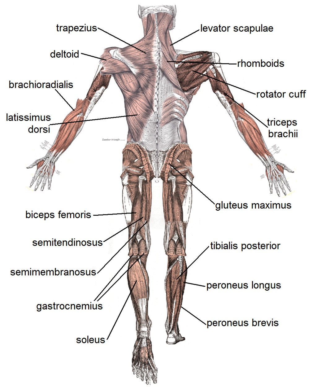
Gastrocnemius (calf muscle): One of the large muscles of the leg, it connects to the heel. It flexes and extends the foot, ankle, and knee. Soleus: This muscle extends from the back of the.
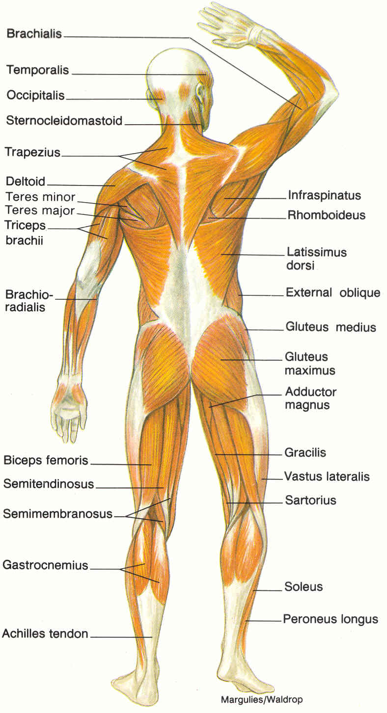
diagram of muscular system Biological Science Picture Directory
Osmosis High-Yield Notes. Muscular system anatomy and physiology. Sliding filament model of muscle contraction. Muscle contraction. Neuromuscular junction and motor unit. Osmosis Muscles high-yield notes offers clear overviews with striking illustrations, tables, and diagrams. Make learning more manageable.
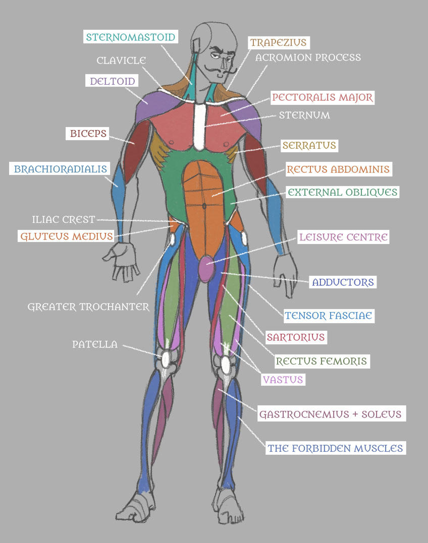
Human Anatomy Muscles with Labels! by Pseudolonewolf on DeviantArt
Gross Anatomy of Skeletal Muscles Head and Neck Muscles Facial Muscles Chewing Muscles Neck Muscles Trunk Muscles Muscles of the Upper Limb Muscles of the Lower Limb Physiology of the Muscular System Skeletal Muscle Activity Nerve Stimulus and the Action Potential Mechanism of Muscle Contraction: The Sliding Filament Theory
Muscles Diagrams Diagram of muscles and anatomy charts HubPages
Muscle diagrams are a great way to get an overview of all of the muscles within a body region. Studying these is an ideal first step before moving onto the more advanced practices of muscle labeling and quizzes. If you're looking for a speedy way to learn muscle anatomy, look no further than our anatomy crash courses .

BodyPartChart Muscular System Front Labeled
Muscular System Anatomy, Diagram & Function | Healthline human body maps muscular system Muscular The primary job of muscles is to move the bones of the skeleton, but muscles also.

Muscles Diagrams Diagram of muscles and anatomy charts
The musculoskeletal system (locomotor system) is a human body system that provides our body with movement, stability, shape, and support. It is subdivided into two broad systems: Muscular system, which includes all types of muscles in the body. Skeletal muscles, in particular, are the ones that act on the body joints to produce movements.
4 human body muscles labeled Biological Science Picture Directory
Each skeletal muscle is an organ that consists of various integrated tissues. These tissues include the skeletal muscle fibers, blood vessels, nerve fibers, and connective tissue. Each skeletal muscle has three layers of connective tissue (called "mysia") that enclose it and provide structure to the muscle as a whole, and also.
Skeletal muscle diagram Healthiack
The coracobrachialis is the smallest of the three muscles that attach to the coracoid process of the scapula. (The other two muscles that attach here are the pectoralis minor and the short head of the biceps brachii.) It is situated at the upper and medial part of the arm. It is supplied by the musculocutaneous nerve.

Muscles Diagrams Diagram of muscles and anatomy charts
Striated muscle; formed of long, multinucleate, unbranched myocytes. Attached at one or either ends to a bony attachment point. Cardiac muscle. Striated muscle; formed of short, uninucleate, branching myocytes which connected at intercalated discs. Specialized muscle of the heart → myocardium. Smooth muscle.
Images 05. Muscular System Basic Human Anatomy
Labelled Diagram of Skeletal Muscle . Skeletal Muscle - Description . The muscle tissue is composed of a large number of myocytes or muscle cells. These muscle cells are slender and long and are termed as muscle fibres. The skeletal muscle fibres are multinucleated. They are arranged parallel to each other, along with some intervening.